How to Extract Experimental Methods from Research Papers with AI (2026)
Linda
Learn how to extract reagents, instruments, and experimental parameters from biomedical papers with AI. Compare real RNA extraction and RT-qPCR methods with citations.
Learn how to extract reagents, instruments, and experimental parameters from biomedical papers with AI. Compare real RNA extraction and RT-qPCR methods with citations.
The Materials and Methods section is where reproducibility lives. A reagent name may appear in one paragraph, an instrument model in another, and the actual temperature or incubation time several pages later. Researchers who compare experimental protocols need all of those details together—without accidentally filling missing parameters from assumptions.
This guide shows how to organize reagents, equipment and experimental settings from published biomedical papers with Noah AI. It follows a source-grounded example involving RNA extraction and reverse-transcription quantitative PCR (RT-qPCR) across three real papers. The goal is a traceable Methods Comparison Table, not an automatically validated laboratory protocol.
On this page
- Key Takeaways
- Why Extracting Methods Entities Matters
- What Makes Methods Extraction Difficult?
- How Noah AI Helps
- Step-by-Step Workflow
- Best Practices & Common Pitfalls
- Example Use Case: RNA Extraction and RT-qPCR
- How to Use Noah AI for Methods Extraction
- Final Checklist
- Conclusion & Next Steps
Key Takeaways
- Experimental methods extraction organizes reagents, instruments, sample inputs, temperatures, times, and controls that are otherwise spread across a paper.
- A useful comparison must preserve each parameter's paper, sample type, experiment arm, Methods subsection, and source citation.
- Methods named-entity extraction can assist review, but a generated table is not proof that omitted details were checked or a protocol is reproducible.
- In this demonstration, Noah organizes reported methods from three published studies involving human skeletal muscle, PBMCs, and lung tissue slices.
- Every extracted setting should be verified in the original Methods text before reuse in an experimental protocol.
Introduction: Why Extracting Methods Entities Matters for Researchers
A methods paragraph can contain several distinct kinds of information. “TRIzol” identifies a reagent; “TissueLyser II” identifies an instrument; “30 Hz” specifies an instrument setting; and “15 min at 12,000 × g” describes a processing condition. These details are related but not interchangeable. Taken out of context, even a correct number can describe the wrong procedural step.
Named entity recognition (NER) is the task of identifying entities such as chemicals, instruments and measurements in text. In a practical research workflow, extraction also requires relation to the experimental context: Which sample was processed? Which arm used the kit? Was a temperature actually reported, or merely suggested by a manufacturer's instructions?
Structured extraction makes it easier to locate details for replication planning, compare reporting across studies, prepare laboratory method inventories, and identify questions for the original authors. It does not replace reading the Methods or the relevant supplementary information. For example, Kuang and colleagues emphasize that qPCR reliability depends on sample processing and quality controls, not simply on naming an extraction kit.([1])
What Makes Methods Extraction Difficult?
Ambiguous terminology. A word like “buffer” may be a reagent category rather than a uniquely identifiable product. Abbreviations such as RT and PCR also need the correct local meaning. A kit name can appear alongside a modification that changes the actual procedure.
Separated experimental details. Sample input may be described under “Sample preparation,” while the reagent appears under “RNA extraction” and the instrument under “Quantitative PCR.” An extractor must connect them without treating different arms or experiments as one continuous procedure.
Units and formatting. The distinction between 1 μg and 1 mg is substantive. OCR or HTML/PDF parsing may damage superscripts, degree symbols, or expressions such as 2 × 10⁶ cells/mL. Centrifugation at 12,000 × g is not automatically equivalent to 12,000 rpm.
Incomplete reporting. Many papers reference manufacturer instructions without providing every pipetting volume, centrifuge model, thermal cycle or catalog number. A responsible extraction records not reported in the article rather than supplying typical laboratory settings.
Comparability. A muscle biopsy, a suspension of peripheral blood mononuclear cells (PBMCs), and pooled human precision-cut lung slices differ in sample matrix and experimental purpose. Their input quantities cannot be ranked as though they came from one standardized experiment.
How AI and Noah Help with Methods Entity Extraction
Rather than manually building a separate notes document for each paper, a researcher can ask Noah to read the relevant full-text Methods sections and organize reported information by category: sample, reagent or kit, instrument, parameter, control and citation. The workflow in this article uses a natural-language research request and a source-linked comparison table.
The practical benefit is organization and triage: the output points the researcher to a specific source section instead of leaving a standalone reagent list with no provenance. It is important to describe this accurately. This case does not demonstrate a dedicated trained NER model, exhaustive PDF extraction, one-click CSV export, or automated validation of experimental reproducibility.
Step-by-Step Workflow for Methods NER
Step 1. Prepare the original Methods sources
Identify papers relevant to the technique and obtain the full text, Methods subsections and available supplements. Specify which experimental arms you want compared. For this example, the research question asks how reagent names, instruments, sample preparation details and settings can be extracted from three biomedical papers on RNA extraction and RT-qPCR.

Figure 1. Research question composed in the Noah Agent input interface. This image shows the question before submission; it is not presented as a completed research-run log.
Step 2. Extract candidate entities with their context
Ask for reagents and kits with their manufacturers, equipment models, sample inputs, experimental conditions and controls. A single entry should represent a specific paper and workflow step. In Kuang et al., for example, the TRIzol extraction and the modified RNeasy kit workflow must remain distinct because they were explicitly compared within the study.([1])
Step 3. Validate against the source text
Check each cell against the original Methods. Preserve published units, record the relevant subsection, and verify that an instrument model or temperature belongs to that experiment. If the full text refers to a manufacturer's protocol without reproducing all settings, flag those details as unreported instead of filling them in.
Step 4. Structure the evidence in a comparison table
Use consistent columns—Study / Sample, Workflow Step, Reagent / Kit, Instrument, Experimental Parameters and Source Citation. The table can then serve as an evidence index for subsequent manual review. Do not merge missing fields into a fictional “complete” protocol.
Step 5. Iterate and document unresolved fields
For unclear entries, ask for a shorter table or a separate verification list while instructing the model not to alter values. Review missing reagent volumes, kit variants, controls, and reported or unreported thermocycling settings. Keep an audit trail of the original source and any manual corrections.
Organize biomedical research evidence with Noah AI
Start with a source-linked methods question, then review the extracted details against the original papers.
Get 300 Free Credits — No Credit Card Required
Free Basic plan: 300 credits on signup; no payment required. See current terms on Noah's pricing page.
Best Practices & Common Pitfalls
- Keep sample context attached. A “1 μg RNA” input is not the same as a “1 mL TRIzol” lysis volume. Report what each number measures.
- Distinguish comparison arms. A paper evaluating multiple kits or reverse-transcription reagents may not have one common protocol applicable to every experiment.
- Preserve manufacturer and version where reported. “RNeasy” is not a sufficient substitute for a specific named kit and any documented modifications.
- Use exact evidence locations. A PMID or DOI identifies a paper; a subsection title makes a parameter easier to verify.
- Record absence honestly. “Not reported” and “not independently verified” describe different limitations.
- Do not infer instrument settings. Convert rpm to relative centrifugal force only when the necessary rotor information is known.
- Review controls and replicates. No-template, no-reverse-transcriptase, biological and technical replicates should not be conflated.
- Do not rank cross-paper performance. Different tissues, assay targets and input units mean descriptive methods comparisons are not controlled performance studies.
Example Use Case: Extracting RNA Extraction and RT-qPCR Methods from Three Papers
To demonstrate the workflow, Noah was asked to organize experimental details from three openly available papers: a human skeletal muscle qPCR study, a PBMC RNA extraction and RT-qPCR optimization study, and an ex vivo human lung-slice study. The resulting report retained their distinct sample contexts and Methods subsections rather than combining them into a new protocol.
| Source paper | Biological material | Examples of recorded details | Methods reference |
|---|---|---|---|
| Kuang et al. (2018) | Human skeletal muscle | TRIzol; TissueLyser II; 10–20 mg sample; 2 × 2 min at 30 Hz; 15 min at 12,000 × g in the TRIzol arm. | RNA extraction; PMID 29746477 |
| Browne et al. (2020) | Human PBMCs | MagMAX mirVana and RNeasy kit comparison; 1 × 10⁶ cell input in most extraction arms; QuantStudio 3. | Kit evaluation; PMID 32265908 |
| Pechous et al. (2024) | Human precision-cut lung slices | 3–6 pooled slices; 1 mL TRIzol; 30 min at room temperature; QuantStudio 6 Flex. | RNA isolation and qRT-PCR; PMID 38940558 |
These are examples of documented methods, not recommendations to combine the steps. Kuang's study compares extraction approaches in muscle; Browne evaluates multiple kits and assay components in PBMCs; Pechous reports an RNA-to-qPCR workflow for human lung tissue slices.([1] [2] [3])
The Noah Methods Comparison Table
Figure 2 shows the nine-row output organized around three workflow stages from each paper. Reagent names and instruments are kept beside experimental settings and a citation field, so a reader can return to the original source.
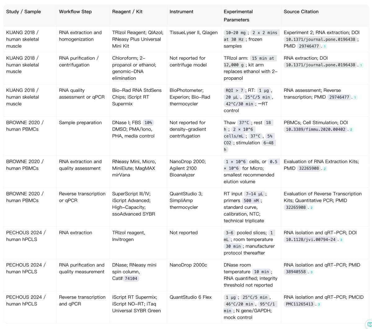
Figure 2. Noah-generated Methods Comparison Table covering reagents, equipment and reported conditions for three published biomedical studies. The screenshot reproduces the research output; values still require source-level verification before laboratory use. On small screens, scroll the image horizontally to view every column.
What the extracted details actually show
Kuang 2018: The study reports frozen human skeletal muscle and different extraction approaches. The TRIzol arm records TissueLyser II homogenization at 30 Hz for two two-minute periods and a 15-minute, 12,000 × g centrifugation step. Its reverse-transcription section reports a 1 μg RNA input in a 20 μL reaction with defined incubation temperatures. Those settings belong to particular procedures; they should not be applied automatically to every arm.([1])
Browne 2020: The paper compares RNeasy Mini, RNeasy Micro and MagMAX mirVana extraction approaches and tests reverse-transcription kits and qPCR components. Sample input depends on the kit, including 0.5 × 10⁶ PBMCs for the Micro condition. It identifies NanoDrop 2000, Agilent 2100 Bioanalyzer and QuantStudio 3, as well as standard curves, calibration material and no-template controls. A kit-comparison value is not automatically a setting in the optimized final assay.([2])
Pechous 2024: Three to six human lung slices were pooled and exposed to 1 mL of TRIzol at room temperature for 30 minutes. The authors also report DNase processing, RNeasy mini-column purification, a NanoDrop 2000c, and a specified reverse-transcription thermal profile before qPCR on QuantStudio 6 Flex. Some qPCR settings are deferred to manufacturer instructions or not stated in the cited subsection; those fields must remain unverified at the article level.([3])
How to Use Noah AI for Methods NER in the Wild
Start in Noah Agent with the original papers or their full-text links. State the biological method to examine and identify the Methods sections that contain the relevant procedures. Then request a structured table with one row per paper and workflow step, including reagent or kit, instrument model, numerical settings and exact source location.
For a compact, verifiable request, a useful instruction is:
“Extract the RNA isolation and RT-qPCR methods from the supplied full-text papers. For each workflow step, record the biological sample, reagent or kit, instrument, reported quantity or setting, and original Methods subsection. Mark inaccessible or unreported parameters explicitly. Do not combine conditions across studies or infer settings from vendor manuals.”
Once the first table is generated, use the original articles to confirm the values most likely to cause downstream errors: concentrations, temperatures, time, centrifugal force, kit names, controls and experimental arms. If you need a shorter screenshot, ask Noah to reformat the existing table without changing the verified values. Do not describe this demonstration as an automated multi-document CSV export unless that workflow has been directly tested.
Final Checklist
- Verify the paper title, DOI and relevant Methods subsection.
- Record the biological sample, condition and experiment arm for each entry.
- Distinguish reagents, kits, instruments and numerical settings.
- Preserve units, temperatures, durations and centrifugation units exactly.
- Identify controls, normalization procedures and replicate types where available.
- Do not fill missing details with typical values or manufacturer defaults.
- Separate observed methods from suggested improvements or replication questions.
- Check supplements and record which documents were accessible.
- Keep source links next to the extracted evidence.
- Have a researcher verify the final table before using it to design an experiment.
Conclusion & Next Steps
Extracting methods information is useful because it turns scattered experimental descriptions into something researchers can inspect, compare and reuse. A source-linked table reduces the effort needed to find a reagent name or instrument setting; it also makes visible the parameters the paper does not provide.
The critical distinction is between organizing what a paper reports and validating what a laboratory should do. The first is an appropriate AI-assisted literature workflow. The second requires the original Methods, supplementary protocols, appropriate expertise and, when necessary, further experimental validation.
Build a Verifiable Experimental Methods Comparison Table with Noah AI
Extract and organize reported reagents, instruments, sample preparation details, and experimental parameters from biomedical research papers.
Use Noah AI to create a source-linked Methods Comparison Table that makes differences between published methods easier to inspect, flags missing information, and supports manual verification against original publications.
The generated table is a research reference, not a validated or ready-to-execute laboratory protocol.